Introduction
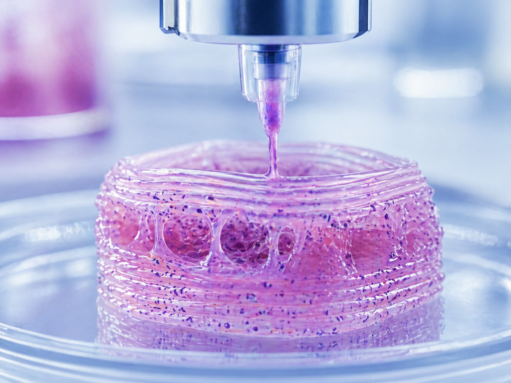
The development of 3D printing methods for synthetic materials, combined with significant advances in biological knowledge, has given rise to the field of 3D biological printing, also known as bioprinting. Unlike traditional two-dimensional cell cultures, where cells grow on flat surfaces and do not accurately reflect the body’s physiological environment, bioprinting enables the creation of three-dimensional structures that replicate the spatial organization of natural tissues. This technology enables the printing of living cells, biocompatible materials, and supporting components to create tissues with functional properties.
Bioprinting has potential for a wide range of applications:
![]() Tissue engineering and regenerative medicine: Bioprinting enables the creation of structures with the potential to replace damaged tissues and even entire organs, thereby reducing dependence on organ donation. As early as the beginning of the 2000s, the first implantation of a printed organ in humans was performed: a urinary bladder produced by researchers at the Wake Forest Institute for Regenerative Medicine, which functioned for more than a decade after implantation. Later, implantation of tissues such as skin, the ear, and the trachea was also reported. However, as of today, organ printing for transplantation remains limited to research and clinical trials, and the regulatory pathway for these products has not yet been fully defined. Experts estimate that it will take several more decades before complex organs such as the heart or lungs reach clinical implementation. By contrast, structurally simpler tissues, such as the cornea, are already at advanced stages of development and even early clinical application.
Tissue engineering and regenerative medicine: Bioprinting enables the creation of structures with the potential to replace damaged tissues and even entire organs, thereby reducing dependence on organ donation. As early as the beginning of the 2000s, the first implantation of a printed organ in humans was performed: a urinary bladder produced by researchers at the Wake Forest Institute for Regenerative Medicine, which functioned for more than a decade after implantation. Later, implantation of tissues such as skin, the ear, and the trachea was also reported. However, as of today, organ printing for transplantation remains limited to research and clinical trials, and the regulatory pathway for these products has not yet been fully defined. Experts estimate that it will take several more decades before complex organs such as the heart or lungs reach clinical implementation. By contrast, structurally simpler tissues, such as the cornea, are already at advanced stages of development and even early clinical application.
![]() Development of new drugs: Beyond direct clinical applications, bioprinting is a significant tool in drug development. Three-dimensional-printed tissues and organs provide a model that more accurately mimics the human body than two-dimensional cultures. Accordingly, they can be used to assess drug efficacy and safety, reduce animal testing, and accelerate drug development.
Development of new drugs: Beyond direct clinical applications, bioprinting is a significant tool in drug development. Three-dimensional-printed tissues and organs provide a model that more accurately mimics the human body than two-dimensional cultures. Accordingly, they can be used to assess drug efficacy and safety, reduce animal testing, and accelerate drug development.
![]() Personalized medicine: Tissue models based on cells derived from the patient can be produced to tailor treatment to the patient’s disease. This approach is particularly relevant for matching treatments to cancerous tumors.
Personalized medicine: Tissue models based on cells derived from the patient can be produced to tailor treatment to the patient’s disease. This approach is particularly relevant for matching treatments to cancerous tumors.
![]() Research: Printed tissue models enable researchers to study diseases and biological processes in a controlled environment that simulates physiological conditions.
Research: Printed tissue models enable researchers to study diseases and biological processes in a controlled environment that simulates physiological conditions.
![]() Bio-devices: Bioprinting can be used to produce tissues integrated with sensors for real-time monitoring of physiological processes. These systems may serve both research and diagnostic applications.
Bio-devices: Bioprinting can be used to produce tissues integrated with sensors for real-time monitoring of physiological processes. These systems may serve both research and diagnostic applications.
![]() Environmental biotechnology: Three-dimensional structures containing microorganisms such as algae, bacteria, or fungi can be printed to break down pollutants in water and soil. These systems function as living biofilters that can operate over time to purify and restore the environment.
Environmental biotechnology: Three-dimensional structures containing microorganisms such as algae, bacteria, or fungi can be printed to break down pollutants in water and soil. These systems function as living biofilters that can operate over time to purify and restore the environment.
![]() Cultivated food based on living cells: Bioprinting enables the creation of three-dimensional structures from muscle and fat cells grown in the laboratory, to produce food products that mimic meat. This approach seeks to produce animal-based food without the need to raise and slaughter animals, while enabling control over composition and texture.
Cultivated food based on living cells: Bioprinting enables the creation of three-dimensional structures from muscle and fat cells grown in the laboratory, to produce food products that mimic meat. This approach seeks to produce animal-based food without the need to raise and slaughter animals, while enabling control over composition and texture.
This review describes the various printing methods, the characteristics of biological ink, the market potential, and the current and future applications of printed products. The review addresses only 3D printing in which the ink contains living cells from which, following printing, a functional three-dimensional tissue develops that mimics natural tissue.
Technology Components
Bioprinter
A machine like those used for standard 3D printing, adapted for the deposition of bioink containing living cells. There are different types of bioprinters based on their printing technologies, each suited to different materials and applications (described in the printing technologies chapter).
Bioink
A core component of the process, containing living cells within a carrier material designed to preserve their viability, functionality, and ability to proliferate and develop into tissues after printing. The ink formulation is tailored to ensure cell stability and viability throughout the printing process and afterward. In some applications, the carrier material degrades or is removed after printing. The viscosity and flow properties of the ink must be matched to the specific printing method.
Printing nozzle/head
Responsible for the precise deposition of the bioink. Its design is adapted to the printing technology and the viscosity of the ink.
Substrate or support structure
A structure into which the bioink is printed. It provides support during the printing process. The support may be temporary or may form part of the final structure.
Computer-Aided Design (CAD) software
Used to create a digital model of the structure to be printed, from which the printer derives the printing instructions.
Control systems
Include hardware and software that manage the printing process while controlling parameters essential to the process, such as temperature, pressure, and printhead movement.
Alongside these components, a sterile environment is required to prevent contamination that could impair cell viability.
Bioink
Bioink consists of a carrier material containing living cells, together with bioactive additives that support cell growth and tissue development.
The carrier material forms the bioink’s physical body. In most cases, it is a hydrogel, which, on the one hand, enables flow during printing and, on the other, preserves the structure’s shape after printing. In addition, it provides the cells with a supportive environment that helps maintain their viability and function. These materials are generally based on natural polymers, such as alginate and collagen, or synthetic polymers, such as polyethylene glycol.
The bioactive additives include proteins and peptides, for example, RGD peptides, laminin, and fibronectin, which are usually derived from the natural extracellular matrix (ECM). Their role is to interact with cells and promote adhesion, proliferation, migration, and differentiation. In the absence of these components, cells trapped within an inert carrier material do not survive over time and do not develop into tissue.
In certain applications, such as hard-tissue engineering (e.g., bone), the bioink is reinforced with ceramic materials. For this purpose, calcium-containing compounds, such as calcium phosphate or hydroxyapatite, are used, sometimes in combination with structural polymers, such as PCL (Polycaprolactone), to provide stiffness and mechanical support to the printed structure.
The development of bioink requires balancing the engineering requirements of the printing process with the need to preserve cell viability and function. Within this context, several key challenges arise:
- Shear stress: During printing, shear forces are exerted on the cells, mainly as they pass through the printhead. These forces may impair cell viability. There is an inherent tension between the need for high printing precision, which requires small nozzles, and the need to preserve cell integrity, since smaller nozzles increase shear stress.
- Tissue contraction: Printed tissues tend to undergo compression and contraction during the maturation stage, in some cases shrinking to as much as half of their original dimensions. Accordingly, the printing dimensions must be planned to compensate for the expected change.
- Structural stability: Soft or large structures may collapse under their own weight during or after printing. To avoid this, supportive scaffolds can be used, or the structure can be stabilized through crosslinking processes, such as exposure to UV radiation, enzyme addition, temperature changes, or chemical reactions. It is important to ensure that these processes do not impair cell viability. In addition, the literature describes an approach in which the ink is printed into a liquid bath with a matching material density, which provides temporary support to the structure during printing.
- Cell density in the ink: A high cell density is required to create functional tissue. However, increasing cell density increases the risk of printhead clogging and reduced cell viability, mainly due to limited oxygen supply to inner cells. Proposed solutions include the use of spheroids, which are three-dimensional clusters of cells, instead of single cells, and the incorporation of cell-protective additives such as nanocellulose, which maintains a stable microenvironment.
- Tissue fusion: After printing, a biological process takes place in which cells and printed structures merge into continuous tissue. Cell-cell interactions and surface tension drive this process, and it is essential for the formation of an intact structure, especially in systems lacking a supportive scaffold.
Printing Technologies
Several main bioprinting technologies differ in how material is deposited, printing resolution, the types of materials supported, and their effects on cell viability.
Extrusion
Extrusion-based bioprinting is the most common method for tissue fabrication. In this method, the bioink is dispensed as continuous filaments, which are deposited layer by layer and merge to form the final structure. The flow is driven by different mechanical mechanisms, such as a screw, piston, or air pressure, which push the material through a nozzle.
![]() Advantages:
Advantages:
Enables the printing of materials with high viscosity and high cell density.
![]() Limitations:
Limitations:
High viscosity requires high pressure, which can lead to significant shear stress and impair cell viability. As a result, cell survival rates may be lower than with other methods.
![]() The method is used to print a variety of tissues, including bone, cartilage, skin, blood vessels, and muscle.
The method is used to print a variety of tissues, including bone, cartilage, skin, blood vessels, and muscle.
Inkjet Printing
This technology is based on the principles of two-dimensional inkjet printers but uses bioink rather than synthetic ink. Printing is performed through the controlled ejection of tiny droplets, which are built up in layers until a three-dimensional structure is formed.
Droplets are generated using two main mechanisms:
- Thermal – pressure generation through rapid heating. However, this method involves challenges such as nozzle clogging and non-uniform droplet size.
- Piezoelectric – pressure generation through electric voltage.
![]() Advantages:
Advantages:
High printing speed, good resolution, relatively low cost, and the ability to combine multiple materials.
![]() Limitations:
Limitations:
The technology is limited to the use of low-viscosity inks and low-cell concentrations, due to the risk of nozzle clogging and increased shear stress.
![]() The method has been successfully applied in the printing of tissues such as skin and cartilage.
The method has been successfully applied in the printing of tissues such as skin and cartilage.
Laser-Assisted Printing
Laser-assisted bioprinting relies on material transfer via focused laser pulses. This process uses a layered structure consisting of a transparent layer, a laser-absorbing metallic layer, and a bioink layer. When a laser pulse strikes the absorbing layer, localized vaporization occurs, generating a gas bubble that propels droplets of bioink toward a receiving surface. The resolution of the printed structure is affected by the laser wavelength and intensity, as well as the bioink’s viscosity.
![]() Advantages: A nozzle-free, non-contact method that enables the use of inks with high cell density while maintaining relatively good cell viability.
Advantages: A nozzle-free, non-contact method that enables the use of inks with high cell density while maintaining relatively good cell viability.
![]() Limitations: High technological complexity, high cost, difficulty in controlling droplet directionality, and a risk of contamination from the absorbing metallic layer.
Limitations: High technological complexity, high cost, difficulty in controlling droplet directionality, and a risk of contamination from the absorbing metallic layer.
Vat Photopolymerization
This method relies on the selective solidification of light-sensitive bioink via controlled irradiation. Printing is carried out in a vat containing liquid bioink. At the same time, a light source, either UV or visible, is directed precisely to defined areas of the ink, causing local solidification. Through layer-by-layer exposure, accurate three-dimensional structures can be built, while areas not exposed to light remain liquid.
An advanced version of this approach, known as volumetric additive manufacturing, enables the creation of three-dimensional structures by projecting tomographic light from multiple directions. In this method, light energy accumulates within defined volumes of the material, and solidification occurs only in regions where an energy threshold is crossed. Unlike layer-based methods, this approach enables the creation of an entire structure within a short time, without gradual layer-by-layer construction and usually without the need for support structures.
![]() Advantages:
Advantages:
Particularly high resolution and the ability to produce complex structures rapidly.
![]() Limitations:
Limitations:
Cell viability is affected by parameters such as light intensity, wavelength, and exposure time, and therefore, a careful balance is required between printing accuracy and cell preservation.
![]() This method is particularly well-suited for creating complex models for research and drug testing.
This method is particularly well-suited for creating complex models for research and drug testing.
Hybrid Printing Technologies
Embedded Bioprinting
In this method, printing is carried out within a support matrix that provides structural stability during printing and prevents collapse or deformation, especially in soft or complex structures. The printing itself is usually performed using extrusion or photopolymerization.
Printing within a stabilized medium may improve the accuracy and resolution of printed structures. Beyond mechanical support, the support matrix also has a biological role: it can provide a physiologically relevant environment, improve cell viability after printing, and even support specific cellular functions and tissue development.
This method is particularly suitable for creating complex tissue and organ models, including vascular networks and structures that require high precision and structural integrity. The choice of matrix material is critical and is usually based on biocompatible hydrogels that can be selectively removed after printing, as needed.
Multimaterial Bioprinting
Multimaterial bioprinting enables the simultaneous printing of multiple materials and cells to create structures that are more complex in both structure and function than those produced by single-material printing. By combining different materials and cells, it is possible to mimic the composition and architecture of natural tissues and to create distinct regions with different functions within the same structure.
Multimaterial printing can be implemented using a range of printing technologies described above. However, one of the main challenges of this approach is maintaining compatibility among the different materials and controlling their interactions during and after printing.
Market Overview
The 3D bioprinting market is expected to grow significantly in the coming years, driven by technological advances, increasing demand for organ transplantation, and the rise of personalized medicine. As the prevalence of chronic diseases increases and the population ages, transplant waiting lists are growing longer, creating a need for innovative solutions to the shortage of organ donors. Printed tissues may provide a possible response to this challenge. In addition, personalized medicine, in which treatments are tailored to each patient’s genetic and health profile, is gaining increasing importance. The ability to print personalized biological models and tissues supports the development of more precise treatment protocols. Bioprinting also affects the development of new drugs and the testing of their efficacy and safety.
Government initiatives and funding mechanisms focused on biotechnology are driving further market development. Efforts to improve the properties of bioink and the printing processes are creating opportunities for the development of advanced products. There is also a growing trend toward integrating artificial intelligence, which simplifies design and technology transfer to manufacturing.
A market survey published in January 2025 projects that the 3D bioprinting market will reach approximately $5 billion by 2032, with a compound annual growth rate of 17% between 2025 and 2032. Other surveys estimate a higher market value, but the differences stem from different definitions of the bioprinting field, some of which also include the printing of synthetic materials for medical purposes.
Despite impressive growth projections, the market has grown more slowly than the forecasts issued 10 years ago. Bioprinting is technologically complex, and its processes are lengthy. Mass production is unlikely in the coming years, and we are still far from printing complex organs. Even when technology becomes more accessible, it is likely to be applied mainly in tissue engineering rather than in the printing of whole organs. More accessible applications today include synthetic skin, ears, bladder, and cornea, which contain a limited number of cell types.
In the food sector, there is a parallel trend of 3D printing to produce cultivated food. This market was estimated at $297 million in 2023 and is expected to reach approximately $2 billion by 2030, with a compound annual growth rate of 34%. However, most food printing involves components that are not living cells, for example, plant-based ingredients or cellular biomass processed before printing.
A small number of companies have attempted to print food from living cells, such as muscle, fat, or connective tissue, to create structures that mimic the texture, taste, and appearance of real cuts of meat. Production costs for this process are likely too high for a food product. In the absence of precise data on the market for cultivated food printing produced from living cells, the assessment is that this market segment currently represents only a very small share of the market, likely less than 1% of all 3D food printing.
Segmentation of the bioprinting market indicates the following main trends:
- By technology: Extrusion printers accounted for the largest market segment in 2025.
- By components: The printer segment was the largest.
- By applications: Regenerative medicine and tissue engineering constitute the main market segment, while personalized medicine applications are expected to grow at the fastest rate.
- By end users: Academic institutions and research institutes are the primary users.
- By geography: North America is the largest market, while the Asia-Pacific region is expected to grow at the fastest rate.
Leading Companies in the Global Market
Manufacturers of bioprinters and bioinks:
| Advanced Solutions Life Sciences, LLC (USA) | CELLINK (BICO Group, Sweden) |
| REGEMAT 3D, SL (Spain) | REGENHU (Switzerland) |
| 3D SYSTEMS (USA) | Poietis (France) |
| Nanoscribe (Germany) | Cyfuse Biomedical K.K. (Japan) |
Companies developing printed tissues:
| Organovo Holdings (USA) | Aspect Biosystems (Canada) |
Companies in Israel
PreciseBio 3D – The company produces tissues from human cells and natural materials for indications in which there is a shortage of organ donors, or to address a medical condition through tissue replacement. The company’s first product is an engineered cornea containing human endothelial cells and human collagen, intended for transplantation in patients suffering from corneal edema. The implant is based on a supportive collagen layer onto which the cells are 3D-printed.
The company faces a unique regulatory challenge because existing regulations are not yet fully adapted to printed biological products, especially those containing living cells. In November 2025, the first-ever human corneal transplantation by the company, created from human cells and printed using unique 3D technology, was performed at Rambam Health Care Campus.
The surgery, performed on a woman in her seventies who had lost sight in one eye due to a corneal disease, was successful: the woman’s vision improved, and the medical team continues to monitor the recovery process.
Challenges to Broad Implementation
Bioprinting is considered one of the most promising technologies in medicine, but several technical, ethical, and regulatory challenges are delaying its broader implementation.
Technological and Biological Challenges
- Vascularization (blood supply): This is one of the central obstacles in the field. Tissues require a dense, complex network of blood vessels to supply oxygen and nutrients to inner cells and remove waste. Existing printing technologies still struggle to create a precise, functional capillary network, and necrosis can develop in the center of the printed tissue.
- Structural complexity: Real organs are composed of a vast range of cell types arranged in a highly precise architecture. Replicating the microscopic structure and the precise interactions among different cell types, such as nerve cells, muscle cells, and connective tissue, is a complex challenge that current technology is still far from fully solving.
- Bioinks: There is a challenge in identifying materials that are both printable, meaning they have suitable viscosity, and capable of providing an ideal environment for maintaining long-term cell viability. In many cases, there is a trade-off between the stability of the printed structure and its suitability for cell growth and development.
Regulatory and Safety Hurdles
- Regulation: Existing pathways are designed mainly for implants that do not contain living tissue. For printed biological products containing living cells, the regulatory pathway remains unclear and undefined.
- Long-term safety: The long-term implications of implanting printed organs are still unknown. Open questions include whether the body may recognize the tissue as foreign after years, whether the components of the bioink are safe for prolonged use, and whether there is a risk of tumor formation from the printed cells.
Ethical and Social Challenges
- Access and equity: Bioprinting is expected to be an expensive technology, especially in its early stages. There is concern that it will be available mainly to wealthier populations, which could deepen gaps in life expectancy and quality of life across different population groups.
- Human enhancement: Future use of the technology to enhance biological performance, such as printing stronger muscles or more durable bones, may blur the line between medical treatment and biological enhancement.
- Cell source: The use of stem cells, and in some cases embryonic cells, raises ethical questions, especially in the context of large-scale tissue production. These issues are not unique to bioprinting but are relevant to any technology that uses such cells.
Summary
3D bioprinting is an innovative technology with broad potential for medical applications, but it is still in the research and development stage. At present, the most common subfield focuses on printing miniature three-dimensional models of human tissues, such as organ-on-a-chip systems or cancer models. These models make it possible to recreate physiological complexity, thereby supporting disease and drug research, reducing the need for animal testing, and enabling the development of personalized treatments. This subfield is easier to implement than printing tissues for transplantation, since it does not require long-term survival or integration into the body, and it is not subject to similarly stringent regulatory requirements.
In the field of regenerative medicine, which focuses on the printing of tissues and organs for transplantation, there is at times an imbalance between media promises and scientific achievements. Despite headlines about success in printing an entire heart, the reality is that progress remains limited to relatively simple tissues – skin, cartilage, or cornea – and even these are not yet in widespread clinical use. The technological and biological challenges are substantial, and the transition from the laboratory to the clinic requires significant breakthroughs in both biology and materials engineering, beyond the printing technology itself. The number of companies active in this field is small. Still, it is worth noting the exceptional achievement of the Israeli company PreciseBio 3D, which has reached a human clinical trial with a printed biological cornea made from living cells.
Applications of the technology in non-medical fields, such as food, remain marginal at this stage. Attempts by a small number of companies, for example, Steakholder Foods in Israel, to produce cultivated food from living cells have been suspended or canceled, most likely because production costs are too high relative to the product’s commercial value.
The information presented on external websites referenced in this report is the sole responsibility of those websites, and the Israel Innovation Authority bears no responsibility for it.
Third parties provided some of this article’s details, information, and data; the Authority does not commit to their accuracy.
This article contains links to external websites. The Authority has no control over the information, details, or data presented on these sites, and such references should not be interpreted as a recommendation, endorsement, or preference for the content, information, or services published on them. The Authority assumes no responsibility for the content of these websites or reliance on the information they provide.